During the delivery, The baby's entire body must pass through the mother's pelvis and the vaginal canal. As we know, the baby's head is proportionally the largest part of his body at this stage, with a diameter equal to that of the thorax.
Due to its size, it is not easy for it to pass through the maternal pelvis, however, nature is very wise and during delivery, the baby's head is adjusted to facilitate its expulsion. And now, thanks to a new study, We can know through three-dimensional images how the baby's head is deformed during childbirth.
Published in the magazine Plos One and performed in France by French and American researchers, the study was performed using magnetic resonance imaging to obtain three-dimensional images that show how the baby's head is deformed to facilitate delivery.
 In Babies and more, how can that head go through the birth canal?
In order to observe how this transformation happens, the researchers they asked seven pregnant women to have an MRI before and during the second stage of their labor: active dilation, which is when the baby's head begins to appear.
After taking the images during the MRI, each woman was immediately transferred to the hospital's maternity wing to continue her labor. The images, show how babies' skull and brains are crushed and deformed to be able to pass through the birth canal.
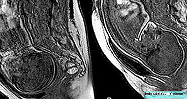
 Three-dimensional reconstruction of the cranial bones before labor (upper images A, C, E) and during the second stage of labor (lower images B, D, F) in patient 5.
 In this first image, the frontal bones are marked in pink, the parietals in green and the occipital bone in blue. In the previous view (A, B), it was observed that the anterior fontanelle decreased in size during delivery. In the side view (C, D), the bones of the skull change in orientation and overlap. In the top view (E, F), it was observed that the anterior and posterior fontanelles decreased in size during the delivery.
 Three-dimensional reconstruction of the fetal brain during MRI before labor (upper images A, C, E) and during the second stage of labor (lower images B, D, F).
 In this second image, it was observed that the brain of all babies changed shape in the second stage of childbirth. This change in the shape of the fetal brain reflected the displacement of the bones of the fetal skull, observed during the second phase of labor.
As the researchers expected, the head of each of the seven babies changed shape to fit the birth canal of their mothers. Of all of them, five recovered the normal form of their heads quickly, while two others remained deformed for a longer period of time, although this had no observable effect on their health.
All children were born healthy, with the exception of one of them, since their results on the Apgar test were lower than normal when they were taken immediately after delivery, however, when taken again 10 minutes later their results were They showed normal. Coincidentally, that baby was one of the three babies who had major deformations in the head during childbirth.

The deformation of each baby's skull depends on several factors, such as the shape of the mother's birth canal, the size of the baby's head and the force with which the tissues hold each section of the skull together.
Some deformation is necessary so that babies can be expelled during childbirth, however, excessive deformation could be a sign that something is wrong, as happened in the case of the baby whose first results of the Apgar test were not normal.
 In Babies and more The baby's head at birth
Because the study was small, the researchers comment that it is necessary to continue researching on this subject to obtain more statistical data, however, this is a good start and could help doctors predict if a mother will have difficulties during childbirth or if the baby is at risk.